Our Office
-
Room 416, 77 Cheongam-ro, Nam-gu, Pohang, Gyeongbuk, 37673, S.Korea
Photoacoustic (PA, also called optoacoustic) imaging combines the strengths of optical and ultrasound techniques to reveal detailed information about biological tissues. Here’s how it works:
Light pulses from a laser are delivered into the tissue.
Absorption of this light by key biological components – such as hemoglobin – causes rapid thermoelastic expansion.
This expansion creates ultrasound waves that travel through the tissue
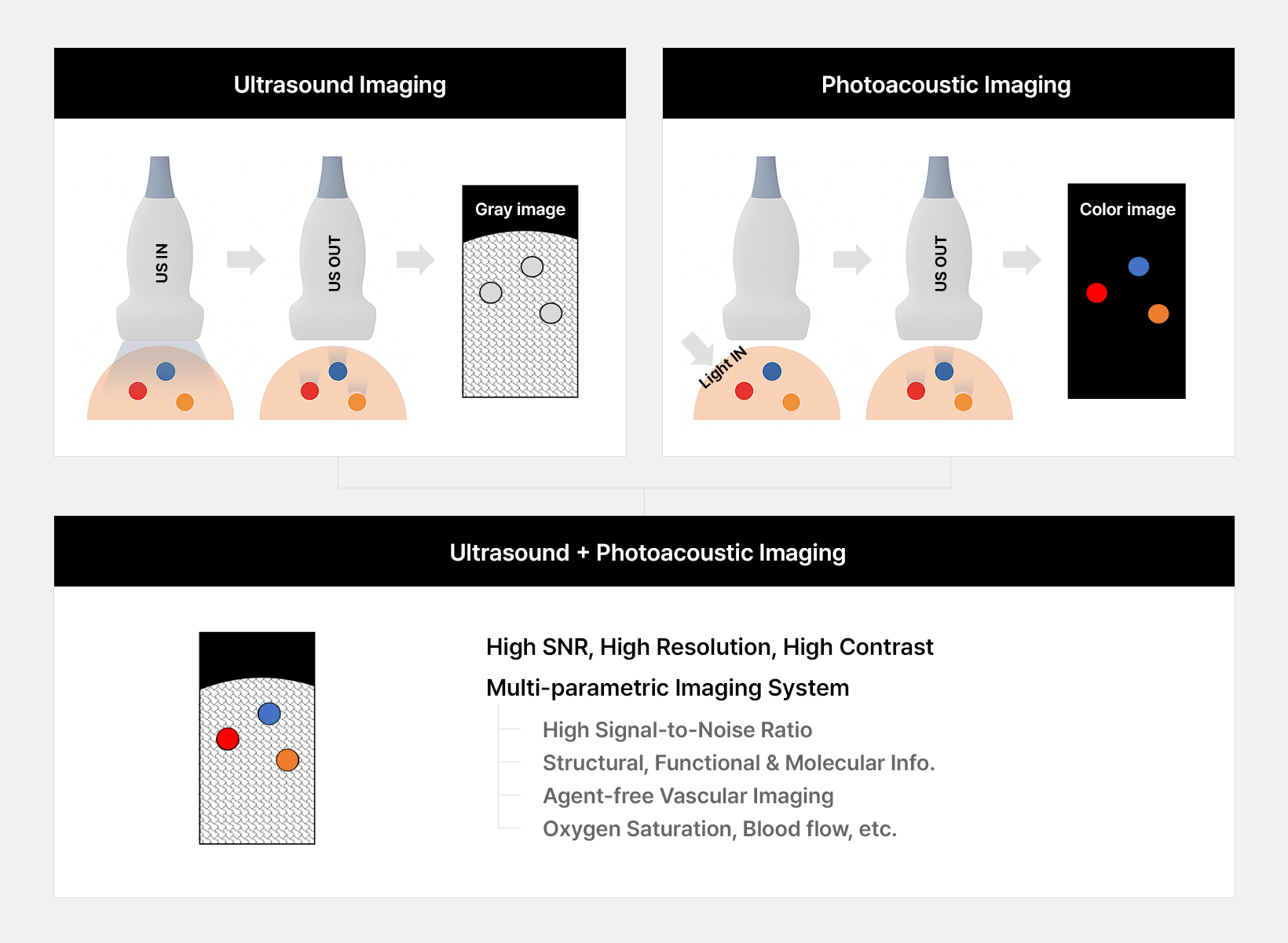
An ultrasound detector captures these waves, and the system reconstructs images that reflect the tissue’s optical absorption properties.
By using ultrasound detection, PA imaging achieves greater imaging depth than purely optical imaging devices. In addition, because intrinsic molecules like hemoglobin absorb light differently at various wavelengths, PA imaging provides wavelength-dependent contrast—enabling visualization of diverse tissue components and physiological features without the need for external contrast agents or invasive procedures. By analyzing spectral information from multiple wavelengths, PA imaging can also estimate parameters such as oxygen saturation.
Deep penetration (several centimeter into tissue)
High spatial resolution
No need for external agents
Real time, non-invasive, and radiation-free
Fusion of light & sound creating new value
© 2024 Opticho. All rights reserved.