Our Office
-
Room 416, 77 Cheongam-ro, Nam-gu, Pohang, Gyeongbuk, 37673, S.Korea
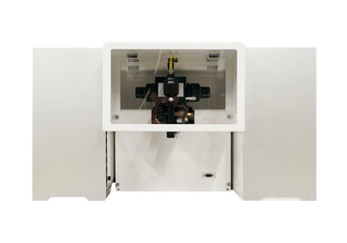
Photoacoustic dermatological imaging device
Photoacoustic microscopy (PAM) is a powerful imaging technique that visualizes optically sensitive structures deep within biological tissues. In both preclinical and clinical settings, real-time imaging is crucial for detecting specific disease markers. However, conventional PAM systems are often limited by their slow motorized scanning stages.
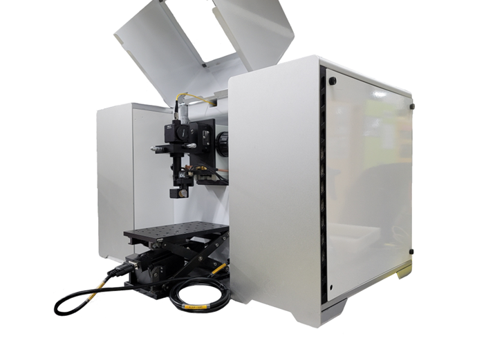
OptichoM introduces a high-speed, high-SNR PAM system powered by an advanced photoacoustic scanner. Its ability to scan in water enables simultaneous transmission and reflection of both ultrasound and laser signals, enhancing imaging efficiency. The system confocally aligns the laser and ultrasound beams to rapidly scan samples. When combined with a motorized stage, this hybrid PAM system achieves a wide scanning range with high-speed acquisition in both optical resolution (OR) and acoustic resolution (AR) modes.
In addition, we offer customizable systems that can be integrated with other optical and acoustic imaging modalities, such as ultrasound microscopy, OCT, and fluorescence imaging.
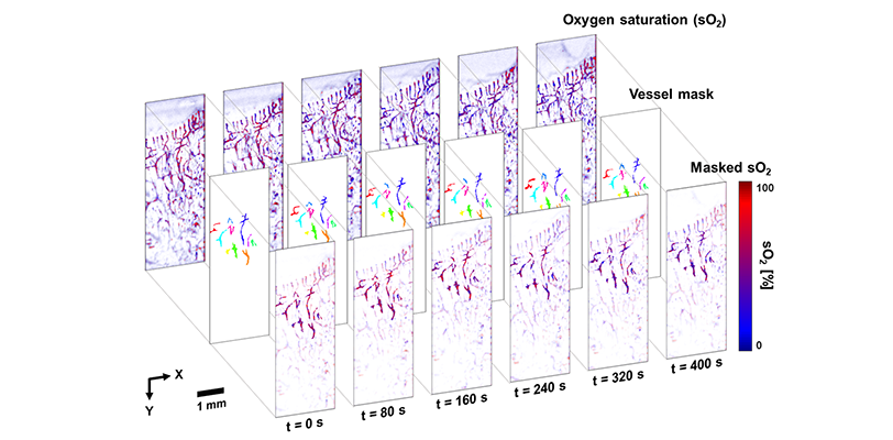
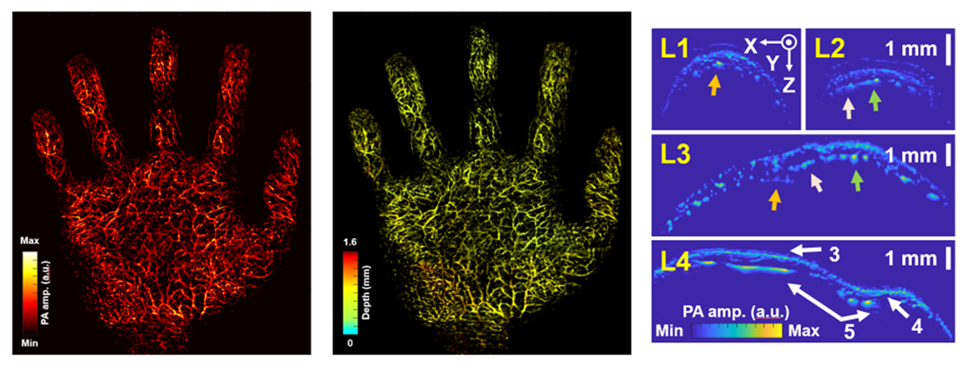
Vascular structural imaging, hemoglobin oxygen saturation
Tumor angiogenic or anti-angiogenic drug response
Tumor progression and regression monitoring
Ischemic diseases
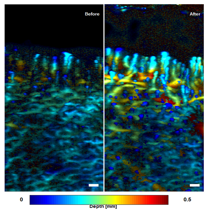
Dermatological diseases
Structural and functional neuroimaging
Ocular imaging
Regenerative medicine
Label-free histology
Molecular imaging using contrast agents
Switchable optical and acoustic resolution modes
Ultrasound microscopy mode
High-speed imaging capability
Excellent signal-to-noise ratio
Wide field of view imaging
Optimized user interface
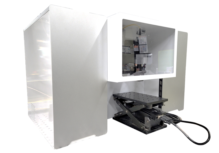
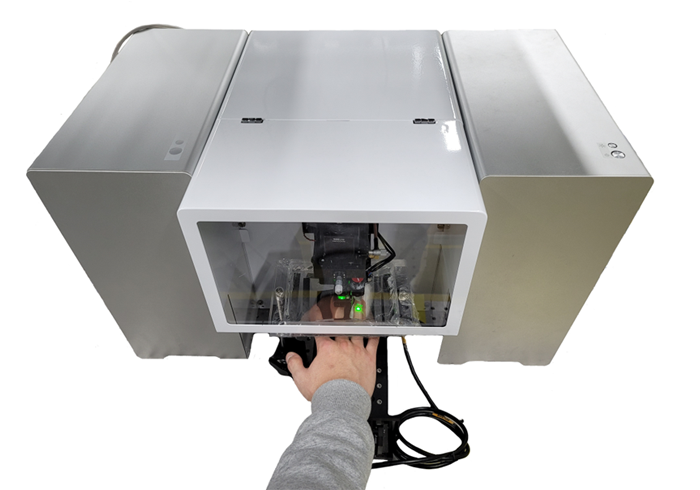
Compact system size
Suitable for both animal and human experiments
Custom-built and multimodal systems are available (OCT, fluorescence, etc.)
| Optical resolution | Acoustic resolution | |
|---|---|---|
| Lateral resolution | ~2.5 - 10 μm | ~80 - 150 μm |
| Axial resolution | ~2.5 - 10 μm | |
| B-scan speed1 | 1-400 Hz | |
| Field of view2 | ~ 30 x 30 mm² + More | |
| Penetration depth | ~ 1 mm in live animals ~ 0.5 mm in humans |
~ 2.5 mm in live animals ~ 2 mm in humans |
| Applications | Animals and humans | |
| Accessories |
|
|

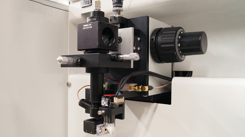
Water-immersible PA galvanometer scanner
Linear motorized stages
Ring US transducer (Fc: 20 MHz)
Opto-acoustic alignment module
Digitizer (12bit, 500 MHz)
Pulse laser
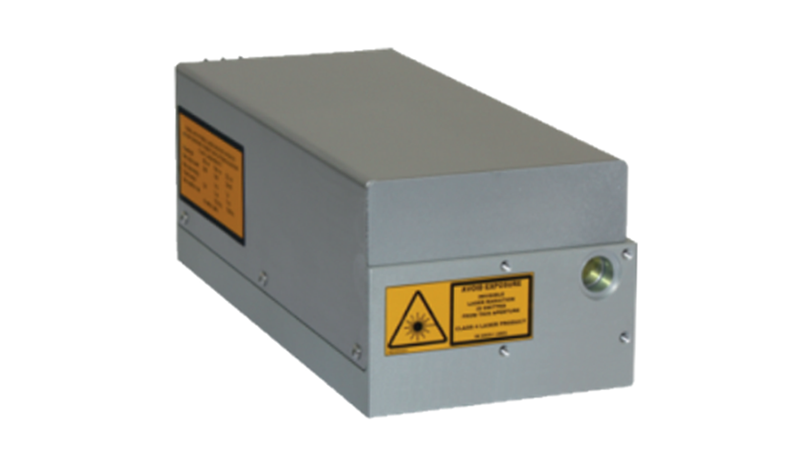
| Pulse laser | |
|---|---|
| Wavelength | 532 nm |
| Pulse repetition rate | Single shot to 100 kHz (Best at 50 kHz) |
| Output power | 5W |
| Pulse width | 2 ~ 10 ns |
| Beam quality (M2) | <1.6 |
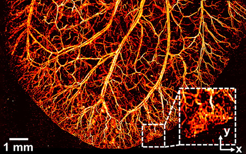
Mouse ear
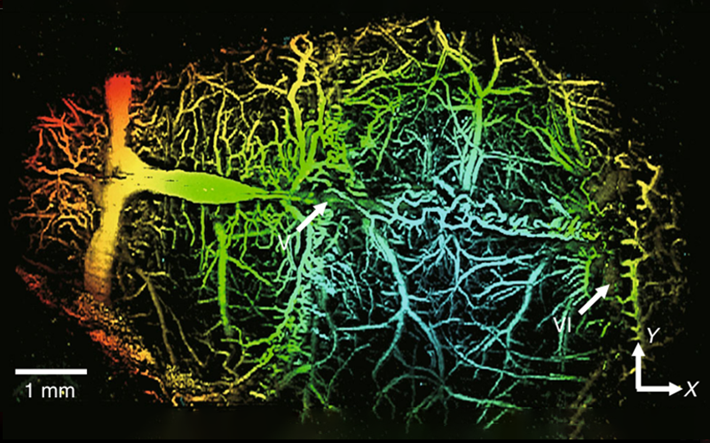
Mouse brain
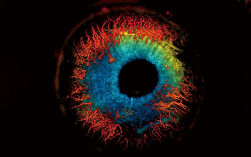
Mouse eye
J. Kim, et al., Light Science & Applications, Vol. 8, 103 (2019)
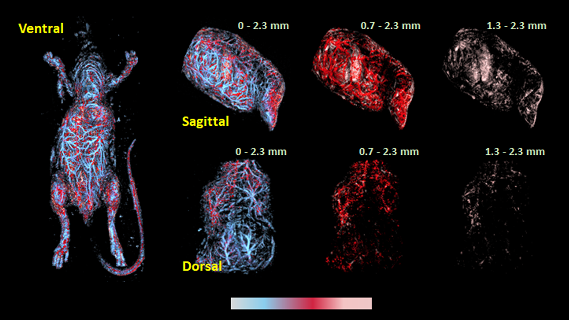
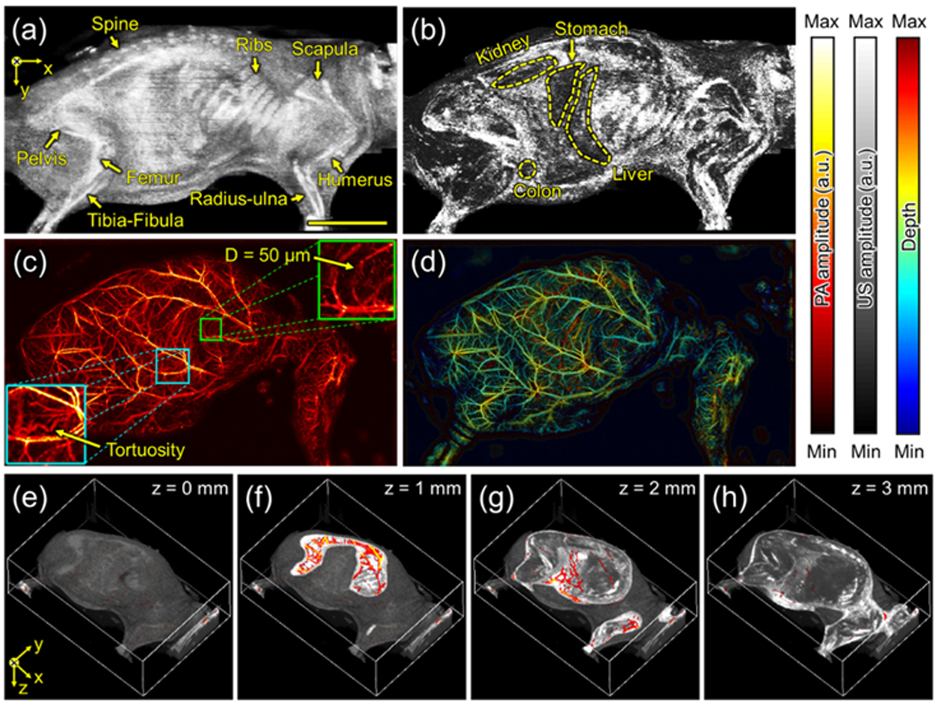
J. Park, et al., PNAS 118, e1920879118 (2021)
J. Ahn, et al., Photoacoustics Vol. 23, 100282 (2021)
J. W. Baik et al., IEEE Transactions on Medical Imaging Vol 39, 975 (2020)
J. W. Baik et al., IEEE Transactions on Medical Imaging Vol 39, 975 (2020)
J. Ahn et al., Biosensors Vol 15, 200 (2025)
Fusion of light & sound creating new value
© 2024 Opticho. All rights reserved.