Our Office
-
Room 416, 77 Cheongam-ro, Nam-gu, Pohang, Gyeongbuk, 37673, S.Korea
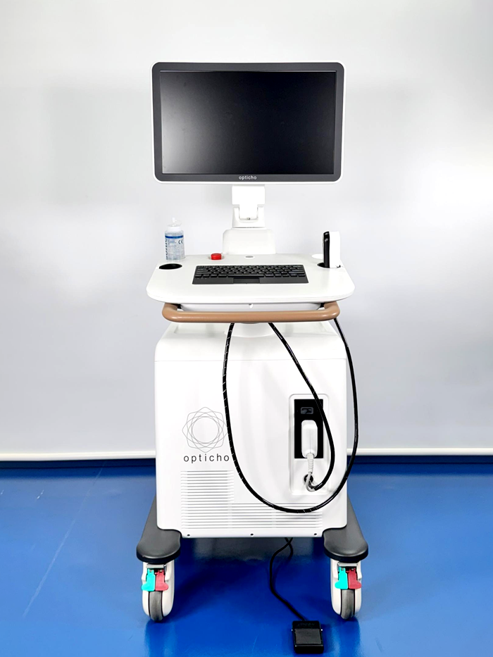
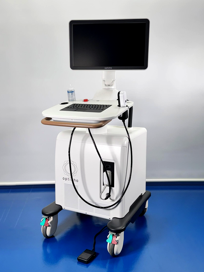
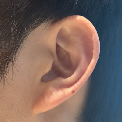
Photoacoustic dermatological imaging device
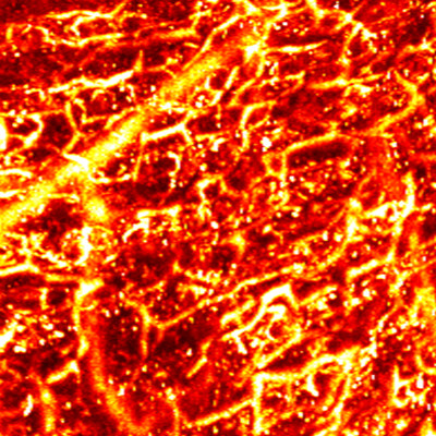
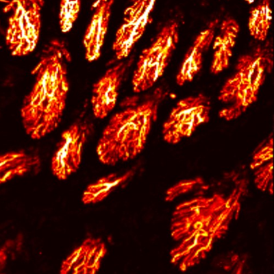
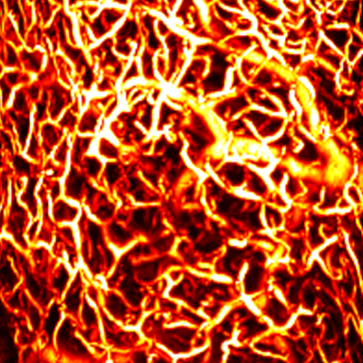
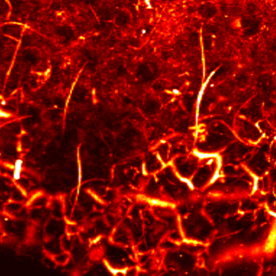
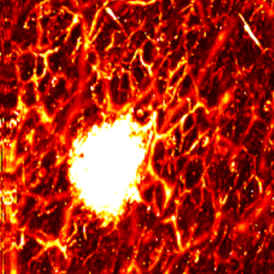
The Dermagio imaging device supports dermatological diagnosis, treatment monitoring, and research. Utilizing advanced photoacoustic technology, Dermagio visualizes microvessels beneath the skin and segments them into anatomical layers—offering structural and functional insights that go beyond traditional imaging methods.
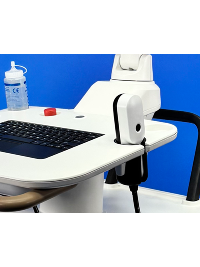
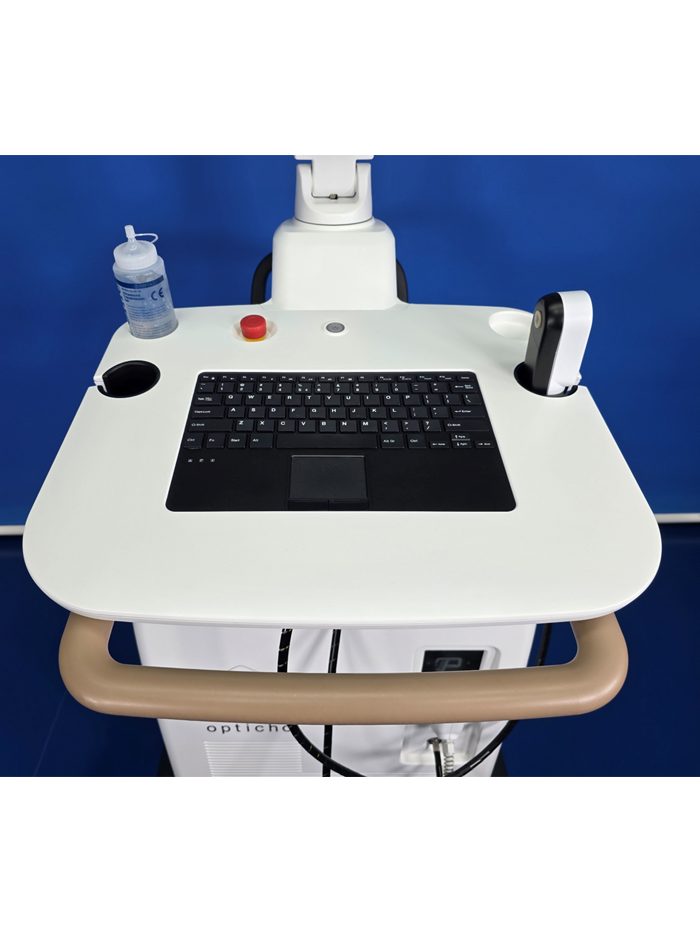
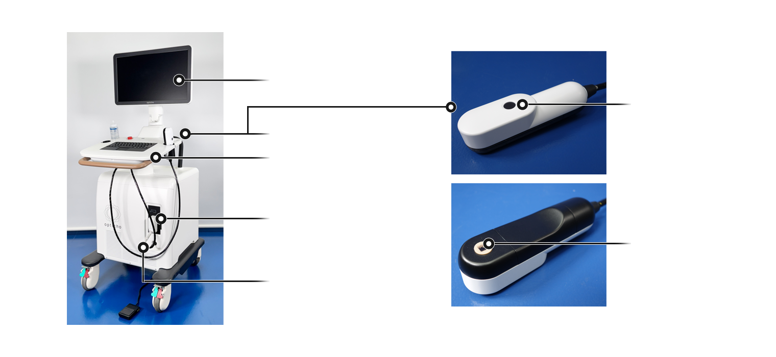
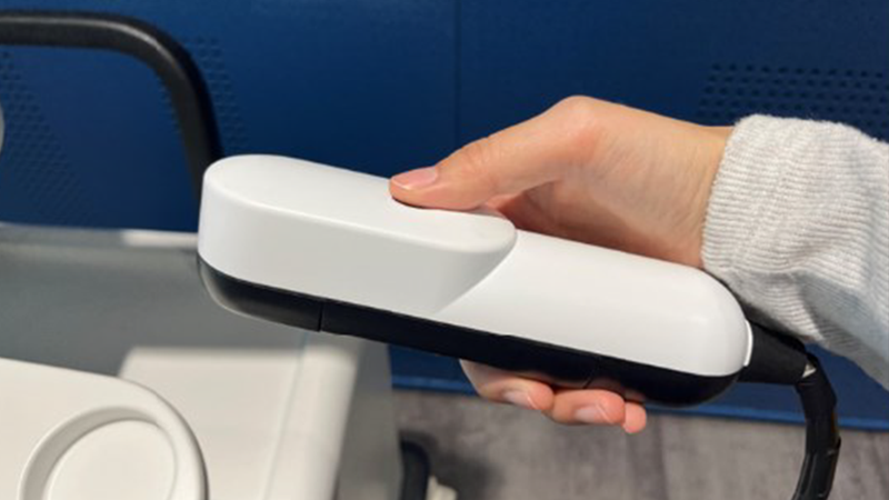
Dermagio consists of three core components: a laser subsystem that delivers imaging light, a handheld photoacoustic probe for direct skin contact, and a control unit for system operation and data processing.
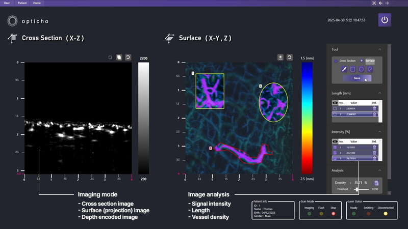
In evaluating skin conditions, Dermagio’s dedicated software enables intuitive visualization and quantitative analysis. Volumetric image data provides depth-resolved information, and skin layers can be segmented based on anatomical structures for independent evaluation. This segmentation also enables advanced applications, such as assessing vasoconstrictive responses or monitoring therapeutic efficacy.
Anatomical layer imaging
Immune and inflammatory skin diseases
Skin cancer (both melanoma and non melanoma)
Vascular conditions and structural imaging
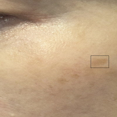
Diagnosis and treatment monitoring of melasma in the epidermis and dermis
Clinical assessment of dermatological agents.
Microneedle research
Skin ageing and skin quality
Tumor progression and regression monitoring
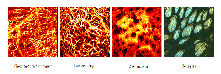
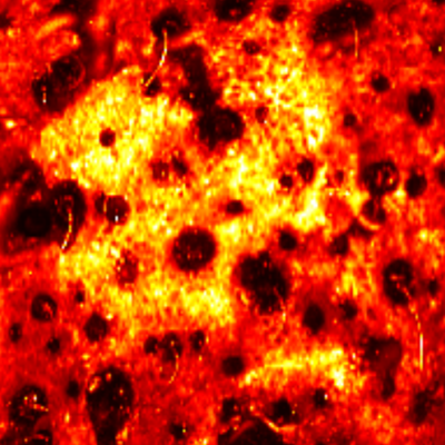
Layer by layer visualization of subdermal microvessels and pigments
Endogenous hemoglobin and melanin allow for daily monitoring
Optics, acoustics, and electronics are configured in one movable unit
A single button on the probe initiates two imaging modes (pre- and standard diagnosis)
Image and quantitative results available immediately after examination
| Spatial resolution |
Lateral resolution (Y-axis): 7.3 μm Axial resolution: 57 μm Elevational resolution (X-axis): 5.0 μm |
|---|---|
| Geometric accuracy |
X-axis: 97.5% Y-axis: 95.5% |
| Imaging speed1 |
Cross section (B-scan): 125 Hz Volumetric imaging time: 1.2 sec for 4 x 4 mm² |
| Sensitivity | 84mV/mM in Evans Blue dye |
| Field of view | ~ 4 x 4 mm² |
| Depth detection | Max imaging depth < 1.8 mm |
| Dynamic range | 15.18 - 47.27 dB |
1 The B-scan speed depends on the step size and pulse repetition rate of the laser.


MAP
Cross sectional view
Surface(Depth) view

MAP
Cross sectional view
Surface(Depth) view

MAP
Cross sectional view
Surface(Depth) view
MAP
Cross sectional view
Surface(Depth) view
MAP
Cross sectional view
Surface(Depth) view
MAP
Cross sectional view
Surface(Depth) view
Fusion of light & sound creating new value
© 2024 Opticho. All rights reserved.